Radiotherapy for brain tumours
What is it?
Radiotherapy can be used in the treatment of brain tumours. Radiation therapy kills brain tumour cells with high-energy x-rays, gamma rays, or protons. It usually follows surgery, although there has recently been a trial looking at using radiotherapy prior to surgery. The radiation kills brain tumour cells that may remain in the area, following surgery. Sometimes, people who can’t have surgery have radiation therapy instead. It may also be given in combination with chemotherapy; this is called combination therapy. Radiotherapy is used in 50% of all cancer patients.
Should I have radiotherapy?
Even if the tumour has been removed, radiotherapy can be recommended to keep hidden cells from growing. This should destroy left over cells and prevent a recurrence of growth. There is a suggestion that, several years after radiotherapy treatment, another brain tumour could develop because of this treatment. There is also a risk of radiation necrosis. Radiation necrosis is a serious, potential late-term complication of radiation therapy. It causes tissue death in the treated area, often months or years after treatment. This is rare. But you should take all factors into consideration. Again, talk with your doctors, trust them and be guided by them. Look at the clinical research evidence. You should not have radiotherapy if you only have a few dividing cells in your cancer. The therapy would not be very successful.
How does radiotherapy work?
Radiation treatments take advantage of the fact that healthy tissue repairs damage in about 8 hours, so that regular low to moderate doses of radiation can be delivered daily. Diseased tissue cannot repair like this, so eventually the tumour cells die as they cannot reproduce effectively. Tumour cells may still show up but they may have lost the power to divide.
The amount delivered to the brain is limited by how much radiation normal tissue can tolerate. Limits are well researched.
Radiation is measured in units called Gray (Gy) or centigray (cGy). The amount of overall dose and amount to be given each day will depend on the tumour type.
How is radiation therapy given?
Little and often. Usually treatment is scheduled Monday to Friday so you will need to pitch up at the hospital every day, with weekends off. Dates and times are usually given in advance and sometimes you can work with the planning team to fit in around your commitments. However, before you get to your first dose there is a significant amount of preparation to be done. This can take a month, and if you have had surgery you will need this time to recover and gather your strength for this treatment. Due to the ‘plastic’ nature of the brain your brain also needs time to recover.
By now you will have met your radiation oncologist. They will discuss your case each week in the MDT meetings. You will need to go to some planning sessions so that all the measurements can be taken. These are called simulations, when the mapping is done to figure out how best to arrange the radiation beams and how best to protect the healthy tissue. If you are having radiotherapy, the chances are that you will need to wear a transparent Perspex mask, made bespoke for you, which is clamped to the table to hold your head in place whilst you are receiving the treatment.
To be honest, having the mask made was worse than the actual treatment for me. The very worse bit? It lasted about one minute. I couldn’t see, I couldn’t move and I couldn’t speak. The straps were being ‘welded’ onto the mask by my ears and it was very hot, burning my ears. I had to wiggle my hands to let the nurse know that something was wrong. After this the whole thing was a breeze.
Patient, Southampton
The mask will enable you to breathe and see normally but it can feel claustrophobic. You will only need to wear it for a few minutes at a time. If you are having whole brain radiotherapy (for secondary brain tumours) you will not need a mask.
Before each session of radiation treatment, you may be asked to put in a mouth guard to protect your gums. Then you will be positioned carefully on a table and the mask fitted. You will then be left alone in the room whilst the kit moves around you, zapping away. It is not in the least painful but you will need to keep still. You may be able to talk to the radiographer who will keep a close eye on you from the next room.
What are the different types of radiation treatment?
Doctors use external and internal types of radiation therapy to treat brain tumours.
External beam radiation therapy
Fractionated external beam radiation therapy (EBRT) is the most common method of radiation therapy used for people with brain tumours. A large machine outside the body aims beams of radiation at the head. Because cancer cells may invade normal tissue around a tumour, the radiation may be aimed at the tumour and nearby brain tissue, or at the entire brain. Some people will also need radiation aimed at the spinal cord.
You will lie on a treatment table in a fixed position. Powerful and precisely placed radiation beams are directed at the tumour using a sophisticated device called a medical linear accelerator (linac). This rotates around you, delivering beams from different angles. When these beams converge on your tumour, the effect is very powerful.
The following details some recent advances in types of radiotherapy. Some of these are standard practice in the UK:
Intensity-modulated radiation therapy (IMRT)
Similar to 3DCRT and is appropriate for both benign and malignant tumours. IMRT uses radiation blocking to sculpt the shape of a ‘dose’ around sensitive structures e.g. the optic nerve. Clinicians can precisely shape the radiation beam so that it conforms to the 3D shape of the targeted tumour. Dose is varied to deliver higher, more effective doses to the tumour, which creates a ‘hot spot’ at the tumour site. It is like 3 to 4 torchlight beams being shone from different directions but at one spot. The light is most intense where the beams meet. But on its own, each beam is relatively weak and passes through normal tissue without much effect. It takes a bit longer to deliver because of the complexity, so about 15 to 45 minutes per treatment. The problem with IMRT is that, with the best will in the world, you move as you breathe. So the very tight margins used by IMRT makes these movements, however slight, a very real problem. This is addressed by using image guided radiotherapy.
Image guided radiotherapy (IGRT)
Prior to the introduction of IMRT, radiation oncologists had to contend with variations in patient positioning, internal movement, and respiratory movement. They do this by treating a wider margin of the tumour. So, for example, to treat a golf ball sized tumour, they would use a tennis ball. With IGRT you can use a golf ball to treat a golf ball.
IGRT gives high resolution, 3D images to pinpoint the tumour site, adjusting your position as necessary, and complete a treatment – all within the time slot.
Proton Beam Therapy
The source of radiation is protons rather than x-rays. This is a type of particle therapy which uses a beam of protons to irradiate the tumour. The doctor aims the proton beam at the tumour. The dose of radiation to normal tissue from a proton beam is less than the dose from an x-ray beam. All protons of a given energy have a certain range; no proton penetrates beyond that distance, so this treatment is good in cases where there is a need for the radiation dose to fall off to zero after it hits the target. In conformal therapy there is exit radiation beyond the tumour, which is why you lose your hair on the side where the beam leaves your head, but proton beams slow down and stop within the target.
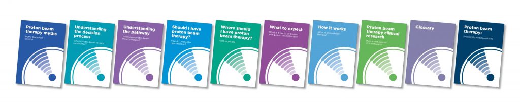
Find our Proton Beam Therapy guides here.
Internal radiation therapy (implant radiation therapy or brachytherapy)
Internal radiation isn’t commonly used for treating brain tumours and is under study. The radiation comes from radioactive material usually contained in very small implants called seeds. The seeds are placed inside the brain and give off radiation for months. They don’t need to be removed once the radiation is gone.
Side effects and useful pointers
During your treatment, you should only use fragrance free and mild soap. So nothing on your hair like gel, or colouring. E45 cream is an excellent moisturizer to use on your skin, which will become burnt.
You will probably not feel any side effects for at least two weeks. Then the following starts to happen:
Fatigue – It is important to stay active. Some people continue to work. However, you must rest when you need to. For more information about fatigue have a look at our fatigue resource.
People warned me that 4 to 6 weeks AFTER the treatment has finished I would feel really tired. This lasted about a month; having a shower was a supreme effort and I had to lie down afterwards. A course of radiotherapy is the equivalent as having another round of major surgery. Listen to your body.
Patient, Leeds
Brain swelling – Sometimes, but not often, radiotherapy causes brain tissue to swell. You may get a headache or feel pressure. Your healthcare team will watch for signs of this problem. They can provide medicine to reduce the discomfort. Radiation sometimes kills healthy brain tissue. Although rare, this side effect can cause headaches, nausea and seizures. Call your doctor immediately if you experience any symptoms that are new or different.
Hair loss – Not everyone will lose their hair. Permanence of hair loss depends on the dose delivered. If there is hair loss, it is gradual. It thins and then becomes patchy, usually where the beams leave your head. Apart from affecting appearance, hair loss makes you notice change in temperatures. Hair will often grow back; it might have a different texture, but you won’t be bald forever.
Skin irritation – Your scalp may become red, dry and tender. Apply moisturiser at any time during your treatment. It is important that you don’t use thick layers. Your healthcare team can suggest ways to relieve these problems.
Nausea – You may be sick or feel sick, particularly if the beams are catching your ear. Again, don’t suffer in silence. Anti-sickness medication will bring relief. Ear congestion – If the beams are passing through your ear, this will become dry and then irritated towards the end and beyond your treatment. You may find that your ear is ‘leaking’. Again, see your doctor, who will provide eardrops.
Weight loss – You may lose weight. This is probably the only time when you will be told it is fine to eat foods that are high in fat. Eat little and often.
Late effects – There is increasing evidence that radiation can cause effects years after treatment, called late effects. These effects can include impairment in memory, concentration and higher mental functions. This is the reason why, wherever possible, focused treatment, particularly radiosurgery (see below), is increasingly used.
Radiosurgery
What is Radiosurgery?
Radiosurgery is a medical procedure used for brain tumour treatment, as well as treatment for other forms of cancer. It uses non-invasive, highly precise radiation beams, usually in one single session, to destroy painlessly or shrink tumours that could otherwise be inaccessible for open surgery.
It is also known as stereotactic radiosurgery (SRS) when used to target brain tumours.
Radiosurgery is effective on discrete, well encapsulated and quite small brain tumours (less then 3 to 4 cm in diameter). Super skinny beams of x-rays or gamma rays are directed at the tumour from different angles. For this procedure, you wear a rigid head frame. The therapy may be given during a single visit (stereotactic radiosurgery) or over several visits. You may also hear these being referred to as Gamma Knife and Cyberknife. These are the commercial names, like Hoover is for vacuum cleaners.
There are some differences between the two. The Cyberknife is a low energy linear accelerator and does not require you to be placed in a frame fixed to the skull. It is frameless SRS. The Gamma Knife is good for particularly small targets and is given after a radiation source helmet is placed on your head. There is also Novalis Tx Radiosurgery.
These all offer a similar (but not identical) minimally invasive approach to brain tumour treatment and are available privately and through the NHS.
Don’t be misled by the words surgery and knife. There is no invasive surgery, so no knife. In this type of treatment though the beams converge on a single point – the tumour. There is no margin added.
What is the difference between radiosurgery and radiotherapy?
Radiotherapy also uses non-invasive radiation beams to treat cancer cells and shrink tumours. But unlike radiosurgery, radiotherapy is normally given as a series of short, daily treatments called fractions and not a single session with a higher dose of radiation. How many fractions or daily treatments will depend on the tumour type and fitness of the patient.
How does radiosurgery and radiotherapy treat cancer?
Radiosurgery and radiotherapy use high-energy radiation beams to deliver the prescribed radiation dose directly to tumour cells, causing them to shrink or die. Treatment results, which are visible on follow-up scans, include shrinkage of the tumour or no further tumour growth. Because cell destruction is a lengthy process, it can often take up to six months before the effect of treatment can be determined by doctors.
What type of patient can radiosurgery benefit?
Due to its power and accuracy radiosurgery can give clinicians the ability to treat patients with cancers once considered untreatable and those for whom surgery is not an option, such as tumours deep in the brain.
Of the many different types of brain tumour that people can contract, you are most likely to have radiosurgery if you have an acoustic neuroma that is less than 3cm across. Radiosurgery can also be used for other brain tumours, including small secondary brain tumours and for people who can’t have brain surgery due to other medical conditions.
Specialists don’t recommend radiosurgery for larger brain tumours. It isn’t possible to get the same dose of radiotherapy throughout the treatment area with a large brain tumour.
Radiosurgery may also not be suitable if there are certain nerves running through the treatment area. The nerves could be given too much radiation. This could cause problems such as hearing loss, depending on the role of the affected nerves.
Did this information make you feel more resourced, more confident or more in control?
Date published: 17-05-2009
Last edited: 1704-2025
Due for review: 04-2028