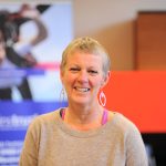
Nikki’s story

In early March 2022, I started to get very bad headaches. They were not like anything I’d experienced before, more like attacks really: very brief – just a minute or two – but intense and disabling. They were always focused in the same place: the left, upper part of my head. It was as if they were trying to tell me something.

I went to my GP, who did some tests: could I stand on one leg? lift both arms? He couldn’t immediately identify a problem. The headaches got worse and in the next few weeks I went to an osteopath, an optician, a pharmacist and A&E. Eventually I went back to my GP and, at the end of April, he sent me for an MRI.

I remember the scan, the claustrophobia and the noise of it. I remember the tiny battered mobile unit in the hospital car park where it took place. I remember the radiographer telling me afterwards that I should not go home. I remember sitting on an uncomfortable trolley behind a blue curtain in A&E for hours. I remember the hot, strong cup of tea someone gave me. I remember a senior nurse, sitting on the end of the bed and holding my gaze as she told me there was a lesion on my brain. What I don’t remember is feeling anything at all. 

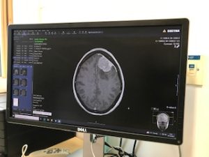
This numbness persisted for the next few weeks, through more scans and phonecalls and appointments and a haze of steroids, as I grew weaker and more debilitated – almost as though the diagnosis had given me permission to let everything go and stop trying. My work, as a freelance writer, ground to a halt.

A letter arrived telling me I’d been referred to a neurosurgeon at a hospital two hours away from my home. He wanted to operate soon. He thought the ‘lesion’ was a large meningioma that had been growing for at least 10 years, maybe 15 or even 20. I thought of all that time it had been there, slowly invading my brainspace. All the things I had done and places I’d been to, unaware of its presence. I wondered how it began? What might possibly have caused it? There are no answers.

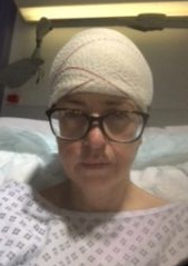
I lost a lot of time when they operated. I recall being driven to a hotel the night before the procedure, but nothing else for three or four days. Apparently, though the initial operation was a success and they removed all the tumour, my brain began to swell immediately afterwards – something they didn’t expect. I had to be transported to a different hospital, in a special for-God’s-sake-don’t-move-her-head wheelchair, for an emergency decompressive craniectomy – the permanent removal of the bone flap to relieve pressure on the brain. I think I was very poorly indeed – though it depends who I ask. My surgeon says ‘you were a bit drowsy’, my mum says ‘I thought I was going to lose you’.

The Glasgow Coma Scale, a measure used by doctors to assess a patient’s level of consciousness, goes from 15 down to three – 15 meaning you’re bright-eyed and bushy-tailed, and three meaning you’re in a deep coma. During those days, my GCS dropped to five. So things could have been worse, but maybe not much.

I finally came round after this second operation, a large piece missing from my skull, my mum and brother sitting by my bed. My memories of the next few days are vague: a woman next to me endlessly repeating the same nonsensical words, a nurse giving me a bed bath at what seemed like four in the morning, a visit from a physio, some tentative walking, a wobbly attempt at stairs. My right side was weak, but functioning, my speech was muddled, but made some kind of sense, my memory was in disarray. They sent me home after about five days. It seemed too soon, but I guess no one really gets better while they are in hospital.

The NHS is chaotic and stretched to its limits, but at the same time it is brilliant. My consultant was highly skilled and made excellent decisions that probably saved my life. I encountered incredible compassion and kindness, from cleaners, porters, health care assistants, nurses. Most doctors. But the NHS doesn’t have the resources for much aftercare or ongoing support. Once I was out of hospital, I felt I was on my own. And sometimes, the isolation and loneliness have been hard to bear.

We usually think about things in a linear way: we say we’re making progress, moving on, taking steps, even going backwards. But my recovery has not been like that. It’s been a ramshackle collection of events that I don’t seem to have much control over.

I’m a single parent and my kids had to go and live with their Dad. I’ve had an absolutely terrifying seizure. I’ve lost my driving licence three times. I’ve had three more surgeries on my skull – one to put a titanium plate in, one to patch up the failing wound, one to take the plate out altogether. (I’m waiting for another op now, to insert a second plate. Fingers crossed.) I worry constantly about money. I have side-effects from my anti-seizure medication – not least mind-warping anxiety.

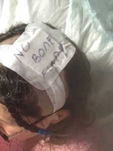
But I am alive, I can walk and talk and even do a bit of work. I am one of the lucky ones. I find humour in bleak places: getting memes from a friend in the midst of awful antibiotic-induced nausea, or taking selfies of the dressing on my scalp on which someone had helpfully written ‘no bone flap’.

I first contacted brainstrust about 18 months after my initial operation. I was desperately low, and I resolved to just ask for help in as many different places as I could. Brainstrust was one of those places and I wish with all my heart someone had told me about them before, that I didn’t have to go through those awful months unsupported. Within days, I had begun coaching sessions with Mariel. Within weeks I had a peer supporter, Imelda – the first person I’d ever spoken to who also had a brain tumour. Within months, I was volunteering to be a peer supporter too. There were so many resources: Facebook groups, Zoom calls, in-person meet-ups. Brainstrust gave me a community, people who know what it’s like to have something go very wrong inside your skull.

I have absolutely no idea what my future holds. I still have terrible days. It’s taken me a week to write this story, when before it would have taken a couple of hours. But I have been able to get perspective and to develop coping strategies. I break tasks down into little chunks, I make lists everywhere, I take one hour at a time. And when things are really bad, I try to find something that makes me laugh. Because without humour, we’re lost, aren’t we?

Introduction

The Brain Tumour Data Dashboard lets you explore up -to-date, population level data about the brain tumours diagnosed in England between 2013 and 2015. Using the drop down menus on the left you can select different groups of patients to view in the charts below. In these charts the number of patients for every 100 diagnoses is displayed as images of people. Patients have been grouped by date of diagnosis, type of tumour, age, gender, and region in England.

For each group of patients you can explore the different routes to diagnosis, the proportion of those who received chemotherapy or radiotherapy, as well as the survival of the patients within each group. For more information about what these metrics mean please see the glossary.

How to use

  1. Select the year of diagnosis using the drop down menu.
  2. Select your patient group of interest from the four drop down menus in the following order:
    1. Tumour group
    2. Age at diagnosis
    3. Region of England
    4. Gender of patient
  3. To view a second chart to compare different groups of patients, click the ‘compare’ button.The second chart will appear below the first chart.

*Note that the tool is best used on a laptop or tablet rather than a mobile phone*

Unavailable data

Some of the data in these charts is not available.There are two main reasons for this:

  1. How the data has been grouped

If you cannot select a patient group from the drop down menus, the data is unavailable because of how the data has been organised.

Public Health England has grouped the data like a branching tree. The bottom of the tree contains all the patients with brain tumours, and then each branch divides the data by a certain characteristics, like age, or location of tumour. But the data is divided in an order, starting with location of the tumour (endocrine or brain), then by age, region, and gender. Age is at the start because it makes a bigger difference to survival rates and treatment rates than gender or region. Sometimes, after the data has been split by type of tumour and age, there is not enough data to be split again. This is because to protect patient confidentiality groups cannot contain less than 100 patients. Because some groups cannot be split further, you cannot create ‘totals’ for everyone by region or gender. For example, you cannot see results for all ages by region, or all brain tumours by gender. If these totals were calculated and released, it might be possible to identify patients, which is why Public Health England cannot release this data.

  1. Statistical reasons and data availability

If you can select a patient group from the chart menus, but the chart does not display, the data is unavailable for one of several reasons:

  1. Data is not yet available for the selected year from Public Health England.
  2. Data is not available because the data quality is too poor to release this statistic.
  3. Data is not available as the statistic is not appropriate for this group.
  4. Data is not available because the standard error of the estimate was greater than 20% and so the estimate has been supressed.

Up to date brain tumour data

Brain tumour data may influence the decisions you make about your care. Data also helps you understand the bigger picture, or landscape, in which you find yourself.

Brain tumour data and statistics influence the focus, and work of organisations like brainstrust. The information helps us to understand the scale and impact of the problems we are setting out to solve.

This tool helps you understand the landscape in which you find yourself having been diagnosed with a brain tumour. This landscape can be particularly tricky to navigate as there are many different types of brain tumour, all of which have a different impact.

The information you see represents the most up-to-date, official, population level brain tumour data available for England. Over time we will be adding to the brain tumour data available and publishing reports, with recommendations, as a result of what we learn from this data.

The data behind this content has come from Public Health England’s National Cancer Registration and Analysis Service (NCRAS) and is a direct result of the ‘Get Data Out’ project.

This project provides anonymised population level brain tumour data for public use in the form of standard output tables, accessible here: http://cancerdata.nhs.uk/standardoutput

Incidence

The number or rate (per head of population) of new cases of a disease diagnosed in a given population during a specified time period (usually a calendar year). The crude rate is the total number of cases divided by the mid-year population, usually expressed per 100,000 population.

Malignant

Malignant tumours which grow by invasion into surrounding tissues and have the ability to metastasise to distant sites

Mortality

The number or rate (per head of population) of deaths in a given population during a specified time period (usually a calendar year). The crude rate is the total number of deaths divided by the mid-year population, usually expressed per 100,000 population.

Non-malignant

Not cancerousNon-malignant tumours may grow larger but do not spread to other parts of the body.

Survival

The length of time from the date of diagnosis for a disease, such as cancer, that patients diagnosed with the disease are still alive. In a clinical trial, measuring the survival is one way to see how well a new treatment works. Also called ‘overall survival’ or ‘OS’.

Routes to Diagnosis

Under the ‘Routes to Diagnosis’ tab in the Brain Tumour Data Dashboard, you can explore the ways patients have been diagnosed with brain tumours. There are many ways, or routes, for cancers to be diagnosed in the NHS. A ‘route to diagnosis’ is the series of events between a patient and the healthcare system that leads to a diagnosis of cancer. The routes include:

  1. Two Week Wait

Patients are urgently referred by their GP for suspected cancer via the Two Week Wait system and are seen by a specialist within 2 weeks where they are diagnosed.

  1. GP referral

Diagnosis via a GP referral includes routine and urgent referrals where the patient was not referred under the Two Week Wait system.

  1. Emergency Presentation

Cancers can be diagnosed via emergency situations such as via A&E, emergency GP referral, emergency transfer or emergency admission.

  1. Outpatient

Outpatient cancer diagnoses include diagnoses via an elective route which started with an outpatient appointment that is either a self-referral or consultant to consultant referral. (It does not include those under the Two Week Wait referral system).

  1. Inpatient elective

Diagnosis via an inpatient elective route is where diagnosis occurs after the patient has been admitted into secondary care from a waiting list, or where the admission is booked or planned.

  1. Death Certificate Only

Diagnoses made by Death Certificate Only are made where there is no more information about the cancer diagnosis other than the cancer related death notifications. The date of diagnosis is the same as that of the date of death.

  1. Unknown

For some patients with a cancer diagnosis, there is no relevant data available to understand the route to diagnosis.


More information

If any of the statistical terms in this section of the brainstrust website are hard to understand, we recommend looking them up here:

Cancer Research UK’s Cancer Statistics Explained

http://www.cancerresearchuk.org/health-professional/cancer-statistics/cancer-stats-explained/statistics-terminology-explained#heading-Seven

If you are looking for help understanding terms relating specifically to brain tumours, and treatment, then the brainstrust glossary is available here:

https://www.brainstrust.org.uk/advice-glossary.php